ZBTB7A
ZBTB7A,全稱為鋅指和BTB結構域包含蛋白7A,是一種含有N端BTB/POZ結構域和C端鋅指結構域的轉錄因子。該蛋白在多種細胞類型中表達,參與基因調控,影響細胞增殖、分化和凋亡等過程。ZBTB7A蛋白通過其鋅指結構域識別特定的DNA序列,結合到靶基因的啟動子區域,從而調控基因表達。研究表明,ZBTB7A蛋白在多種生理和病理過程中發揮重要作用,包括細胞周期調控、細胞分化、細胞凋亡等。ZBTB7A蛋白的表達異常與多種疾病相關,如癌癥、免疫疾病和神經系統疾病等。針對ZBTB7A蛋白的藥物研發正在積極進行中,包括小分子抑制劑、RNA干擾技術和基因編輯技術等。這些藥物有望通過調節ZBTB7A蛋白的表達或活性,從而治療相關疾病。
熱銷產品
ZBTB7A Recombinant Monoclonal Antibody (CSB-RA063930A0HU)
驗證數據
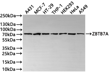
Western Blot
Positive WB detected in: A431 whole cell lysate(30μg), MCF-7 whole cell lysate(30μg), HT-29 whole cell lysate(30μg), THP-1 whole cell lysate(30μg), HEK293 whole cell lysate(30μg), HeLa whole cell lysate(30μg), A549 whole cell lysate(30μg)
All lanes: ZBTB7A antibody at 1:1000
Secondary
Goat polyclonal to rabbit IgG at 1/40000 dilution
Predicted band size: 61 kDa
Observed band size: 70 kDa
Exposure time:2min
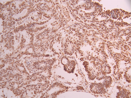
IHC image of CSB-RA063930A0HU diluted at 1:100 and staining in paraffin-embedded human colorectal cancer performed on a Leica BondTM system. After dewaxing and hydration, antigen retrieval was mediated by high pressure in a citrate buffer (pH 6.0). Section was blocked with 10% normal goat serum 30min at RT. Then primary antibody (1% BSA) was incubated at 4°C overnight. The primary is detected by a Goat anti-rabbit polymer IgG labeled by HRP and visualized using 0.05% DAB.
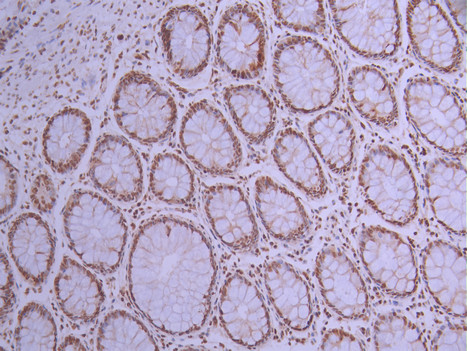
IHC image of CSB-RA063930A0HU diluted at 1:100 and staining in paraffin-embedded human small intestine tissue performed on a Leica BondTM system. After dewaxing and hydration, antigen retrieval was mediated by high pressure in a citrate buffer (pH 6.0). Section was blocked with 10% normal goat serum 30min at RT. Then primary antibody (1% BSA) was incubated at 4°C overnight. The primary is detected by a Goat anti-rabbit polymer IgG labeled by HRP and visualized using 0.05% DAB.
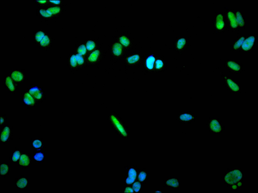
Immunofluorescence staining of MCF-7 cell with CSB-RA063930A0HU at 1:50, counter-stained with DAPI. The cells were fixed in 4% formaldehyde, permeabilized using 0.2% Triton X-100 and blocked in 10% normal Goat Serum. The cells were then incubated with the antibody overnight at 4°C. The secondary antibody was Alexa Fluor 488-congugated AffiniPure Goat Anti-Rabbit IgG(H+L).
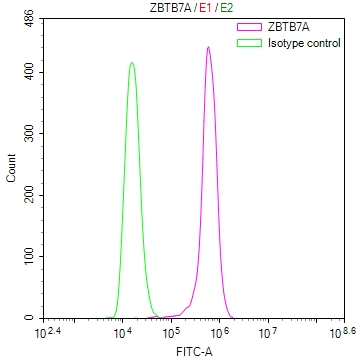
Overlay Peak curve showing BxPC-3 cells stained with CSB-RA063930A0HU (red line) at 1:100. The cells were fixed in 4% formaldehyde and permeated by 0.2% TritonX-100 for?10min. Then 10% normal goat serum to block non-specific protein-protein interactions followed by the antibody (1ug/1*106cells) for 45min at 4℃. The secondary antibody used was FITC-conjugated goat anti-rabbit IgG (H+L) at 1/200 dilution for 35min at 4℃.Control antibody (green line) was Rabbit IgG (1ug/1*106cells) used under the same conditions. Acquisition of >10,000 events was performed.
ZBTB7A Antibodies
ZBTB7A for Homo sapiens (Human)
| 產品貨號 | 產品名稱 | 種屬反應性 | 應用類型 |
|---|---|---|---|
| CSB-PA026343GA01HU | ZBTB7A Antibody | Human,Mouse | ELISA,WB,IHC |
| CSB-RA063930A0HU | ZBTB7A Recombinant Monoclonal Antibody | Human | ELISA, WB, IHC, IF, FC |
ZBTB7A Proteins
ZBTB7A Proteins for Homo sapiens (Human)
| 產品貨號 | 產品名稱 | 來源 |
|---|---|---|
| CSB-YP026343HU CSB-EP026343HU CSB-BP026343HU CSB-MP026343HU CSB-EP026343HU-B |
Recombinant Human Zinc finger and BTB domain-containing protein 7A (ZBTB7A) | Yeast E.coli Baculovirus Mammalian cell In Vivo Biotinylation in E.coli |